What Is a T2 Hyperintense Lesion in the Liver?
 Written By
Abel Tamirat, MD

If you’ve recently had an MRI and were told you have a “T2 hyperintense lesion” in your liver, it’s natural to feel concerned. But take a deep breath — this finding isn’t automatically serious. In fact, many of these lesions are benign and quite common.
In this article, we’ll walk you through what the term means, what causes these liver lesions to appear bright on MRI scans, and how doctors determine whether they’re harmless or need more attention.
Understanding the Term: What Does “T2 Hyperintense” Mean?
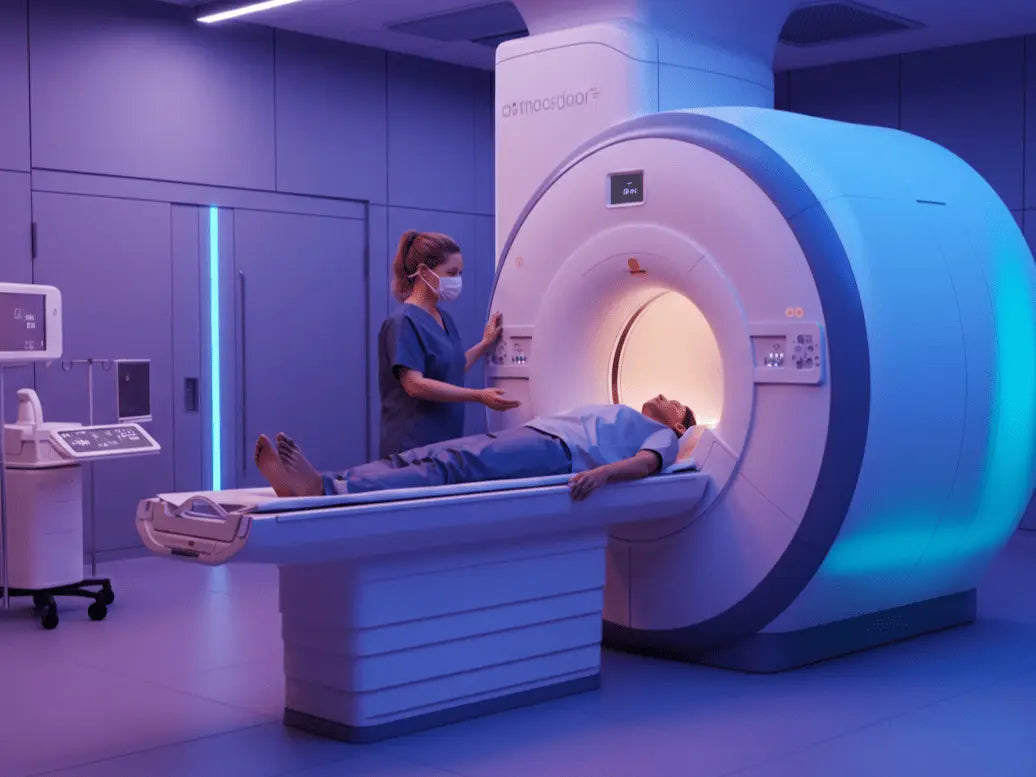
MRI (Magnetic Resonance Imaging) is a type of scan that shows detailed pictures of your internal organs — including your liver. It uses radio waves and magnets to create images. These images can look different depending on how much water, fat, or tissue is in a given area.
T2 refers to one specific setting on the MRI scanner. In a T2-weighted image:
-
Areas with more fluid appear bright (white or light gray).
-
Areas with less fluid appear darker.
So when a report says you have a “T2 hyperintense lesion,” it means there’s a spot in your liver that shows up brighter than the surrounding tissue — usually because it contains more water or blood.
Let’s break that down more clearly:
-
“T2” = the type of image used
-
“Hyperintense” = appears brighter than usual
-
“Lesion” = an area that looks different from normal liver tissue
Curious how different liver textures appear on imaging? This guide explains what is an echogenic liver and how it's interpreted in ultrasound findings.
Why Bright Spots Happen: Common Causes

Seeing a bright spot in your liver on a T2 scan doesn’t necessarily mean cancer or anything life-threatening. There are many harmless reasons a lesion might appear bright, especially if you're not experiencing any symptoms.
Here are the most common types of T2 hyperintense lesions doctors see:
1. Simple Liver Cysts
These are fluid-filled sacs that form in the liver. Most people don’t even know they have them.
-
What they look like: Round, smooth, and very bright on T2 imaging
-
Are they dangerous? No. Simple cysts are usually harmless and don’t require treatment.
Learn what size of liver cyst is dangerous and when your doctor may recommend monitoring or follow-up.
2. Hemangiomas
Hemangiomas are clusters of small blood vessels. They’re the most common benign (non-cancerous) liver tumors.
-
What they look like: Very bright on T2, often with a specific pattern of blood flow when contrast is used
-
Are they dangerous? No. Unless they are very large or cause pain, they usually don’t need treatment.
3. Focal Nodular Hyperplasia (FNH)
This is a benign liver growth made up of normal liver cells. It often occurs in younger adults.
-
What they look like: Slightly bright on T2 with a central scar (which also appears bright)
-
Are they dangerous? Rarely. Most people with FNH live normal, healthy lives without needing surgery.
FNH is often stable, but to understand more serious changes, see this guide to liver cancer stages.
4. Liver Adenomas
Adenomas are benign tumors more common in people assigned female at birth, especially those taking hormonal birth control.
-
What they look like: Varies — they can be slightly bright if they contain fat or blood
-
Are they dangerous? Sometimes. Larger adenomas carry a small risk of bleeding or turning cancerous, so doctors may monitor them more closely.
Hormone-sensitive liver adenomas can be influenced by diet — explore this liver reduction diet to reduce liver fat and pressure.
When a Bright Lesion Could Be Something More
Although many bright lesions on T2 imaging are benign, some types of liver cancer or secondary tumors (called metastases) can also appear bright, especially if they have fluid or dead tissue inside.
These are the signs that usually make doctors more cautious:
-
Irregular shape or borders
-
Heterogeneous appearance (parts of it are bright, others dark)
-
Changes after contrast injection (like rapid wash-in and wash-out)
-
Associated symptoms like weight loss, fatigue, or jaundice
In people with known liver disease or a history of cancer, a bright lesion might prompt further testing — such as a biopsy, follow-up MRI, or CT scan — to be sure of the diagnosis.
If you're managing liver issues and considering advanced care, here’s what to know about what disqualifies you from a liver transplant.
What Does It Mean for You?
If your radiology report mentions a T2 hyperintense lesion, your doctor will consider several factors before making a decision:
-
Your medical history (do you have liver disease or cancer?)
-
Size and shape of the lesion
-
Any symptoms you’re experiencing
-
Results from contrast-enhanced MRI, if done
Sometimes, they may recommend repeating the scan in 6–12 months to make sure nothing changes. Other times, if the lesion looks completely typical (like a cyst or hemangioma), they may say no follow-up is needed at all.
Many lesions turn out to be benign — see liver lesions explained for an in-depth breakdown of types and outcomes.
What Happens Next?

Depending on what the lesion looks like and your overall health, here are a few possible next steps:
1. No Action Needed
If the lesion looks like a simple cyst or classic hemangioma, your doctor may simply reassure you and not recommend any follow-up.
2. Repeat Imaging
If the lesion is unclear or new, your provider might schedule another MRI or ultrasound in a few months to see if anything changes.
3. Contrast-Enhanced MRI
In some cases, doctors use special dyes during the scan to better understand blood flow in the lesion — which helps distinguish between benign and malignant spots.
4. Biopsy or Referral
If there are concerns based on your history or imaging, your doctor may refer you to a liver specialist (hepatologist) or recommend a small tissue sample be taken for lab analysis.
The Role of Other Imaging Tools
Besides T2 imaging, radiologists often look at:
-
T1-weighted images: These can show fat, bleeding, or solid tissue
-
Diffusion-weighted imaging (DWI): Helps tell how dense the tissue is — tumors often restrict diffusion
-
Hepatobiliary phase (using special liver-specific contrast agents): Helps identify lesions like FNH or adenomas more clearly
By combining these techniques, doctors build a full picture of what’s happening inside your liver.
Lesions with poor contrast uptake or fast washout may be more serious — here's how can high liver enzymes be dangerous fits into the bigger diagnostic picture.
You’re Not Alone — And You’re Not Powerless
Liver findings on imaging can sound intimidating, but remember that radiologists read thousands of these scans a year. Most small T2 hyperintense lesions are not dangerous — and even when they need monitoring, there are many effective ways to manage liver health.
If you’re feeling overwhelmed, it’s okay to ask your doctor to walk you through the results in plain language. Don’t be afraid to bring questions. It’s your body, and you deserve to understand what’s going on.
What You Can Do Right Now

Here are a few proactive steps you can take:
-
Keep a copy of your MRI report and ask your provider for a layperson's explanation.
-
Stay on top of your follow-ups, if recommended.
-
Support your liver health by maintaining a balanced diet, avoiding excessive alcohol, and managing any underlying conditions like diabetes or high cholesterol.
And most importantly, try not to jump to conclusions. Imaging findings are just one piece of the puzzle — and they often turn out to be harmless.
Want to take more control over your liver health?
Try Ribbon Checkup’s at-home liver tests—simple, accurate, and convenient.
Related Resources
-
What Size of Liver Cyst is Dangerous? What You Need to Know
Simple cysts are a common cause of T2 hyperintense lesions. Learn when their size or appearance may warrant follow-up. -
Liver Lesions: Causes, Diagnosis, and Treatment Options
Explore the various types of liver lesions, what causes them, and how they're evaluated through imaging like MRI. -
Can Ultrasound Detect Liver Cancer?
When a lesion looks suspicious on MRI, ultrasound is often used as a complementary tool. Learn what it can (and can't) detect.
References
Gatti, M., Maino, C., Tore, D., Carisio, A., Darvizeh, F., Tricarico, E., … Faletti, R. (2022a). Benign focal liver lesions: The role of magnetic resonance imaging. World Journal of Hepatology, 14(5), 923–943. https://doi.org/10.4254/wjh.v14.i5.923
Gatti, M., Maino, C., Tore, D., Carisio, A., Darvizeh, F., Tricarico, E., … Faletti, R. (2022b). Benign focal liver lesions: The role of magnetic resonance imaging. World Journal of Hepatology, 14(5), 923–943. https://doi.org/10.4254/wjh.v14.i5.923
Hiroki Haradome, Grazioli, L., Morone, M., Sebastiana Gambarini, Kwee, T. C., Takahara, T., & Stefano Colagrande. (2012). T2‐weighted and diffusion‐weighted MRI for discriminating benign from malignant focal liver lesions: Diagnostic abilities of single versus combined interpretations. Journal of Magnetic Resonance Imaging, 35(6), 1388–1396. https://doi.org/10.1002/jmri.23573
Liver Lesions: What They Are, Types, Symptoms & Causes. (2023, September 7). Retrieved June 16, 2025, from Cleveland Clinic website: https://my.clevelandclinic.org/health/diseases/14628-liver-lesions
Zimlich, R. (2022, August 16). What Can an MRI of the Liver Detect? Retrieved June 16, 2025, from Healthline website: https://www.healthline.com/health/mri-liver

Dr. Abel Tamirat is a licensed General Practitioner and ECFMG-certified international medical graduate with over three years of experience supporting U.S.-based telehealth and primary care practices. As a freelance medical writer and Virtual Clinical Support Specialist, he blends frontline clinical expertise with a passion for health technology and evidence-based content. He is also a contributor to Continuing Medical Education (CME) programs.


