Can Ultrasound Detect Liver Cancer?

Can an ultrasound scan detect liver cancer? Yes – ultrasound is a commonly used imaging test that can reveal tumors or masses in the liver. Doctors often turn to ultrasound as a first step when liver cancer is suspected, because it’s a quick, noninvasive way to see if any abnormal lesions are present in the liver. However, while ultrasound can detect liver cancer, it’s not foolproof. Very small tumors or certain conditions can sometimes hide liver cancers from ultrasound’s view. In this article, we’ll explore how liver ultrasounds work, their role in early cancer detection, and what to expect if you undergo one. We’ll also compare ultrasounds to other imaging tests (like CT scans and MRIs) and discuss how tools like at-home liver tests can complement your liver health monitoring routine.
Why Liver Cancer Detection Matters Early
Catching liver cancer early can make a life-saving difference. Often called a "silent" disease, liver cancer’s early-stage tumors frequently don’t cause noticeable symptoms. Symptoms like pain or jaundice typically appear when the disease is advanced, making treatment harder. That’s why proactive screening and early detection are crucial.
When confined to the liver, liver cancer is much more treatable. Localized cases have a 5-year survival rate of around 37%, with early-stage surgeries or transplants improving rates to 60–70%. In contrast, advanced liver cancer that spreads to distant organs has a survival rate of only about 3%.
Screening high-risk individuals with ultrasound and blood tests can reduce liver cancer mortality by 37%. Detecting tumors early provides more treatment options and better outcomes, giving patients a fighting chance.
Understanding the different stages of liver cancer can help you grasp why early detection is so critical.
What Is an Ultrasound and How Does It Work?
Ultrasound (or sonography) is an imaging technique that uses sound waves to create pictures of the inside of your body. Unlike CT or X-rays, it involves no radiation. A handheld transducer sends high-frequency sound waves and listens for echoes that bounce back, which are then converted into real-time images by a computer.
-
Think of it like a flashlight reflecting off objects in a dark room. Sound waves bounce off tissue boundaries differently, creating an image.
-
Solid structures, like a liver tumor, reflect sound strongly, while fluid-filled areas allow sound to pass through.
Key facts about ultrasound:
-
Images appear in shades of gray, showing organ texture and abnormalities.
-
No pain – the probe may apply slight pressure, but no needles or incisions.
-
Safe, fast (20–30 minutes), and widely available.
-
Cost-effective compared to CT or MRI.
-
Excellent for real-time imaging of motion, blood flow, and guiding procedures like biopsies.
It’s also frequently used alongside liver enzyme tests for a more complete diagnostic picture.
How Ultrasound Helps Detect Liver Tumors
When it comes to detecting liver tumors, ultrasound is often the first imaging test doctors use. On the screen, a tumor typically shows up as a spot or mass that looks different from the surrounding healthy liver tissue. Depending on its makeup, it might appear darker (hypoechoic) or brighter (hyperechoic). Solid tumors often reflect more sound, showing up as brighter areas, while cystic regions may look darker. The sonographer takes still images and measurements of anything that stands out. Not every spot is cancer—benign cysts, fatty deposits, or scar tissue can look similar. This is especially true in patients with fatty liver or liver lesions, where texture can be misleading.
Learn more about how at-home liver tests work and what they can reveal about your liver health in this comprehensive guide.
Can Ultrasound Spot Early-Stage Liver Cancer?

One key question is how effective ultrasound is at detecting very small, early-stage liver cancers. The answer: it can catch some, but it’s not 100% sensitive for tiny tumors. Research shows that in surveillance settings (screening people with liver cirrhosis for early HCC), ultrasound alone may detect about 63% of early-stage liver cancers, meaning roughly 1 in 3 small tumors could be missed.
Early tumors can be tiny (under 2 cm) and may blend in with the background liver tissue. Certain tumor locations, like high under the diaphragm, are also harder to visualize. To improve detection, doctors often combine ultrasound with AFP blood tests and liver enzyme monitoring.. Regular ultrasounds (every 6 months) further increase the chances of spotting early cancers.
You can also start monitoring your liver enzymes from home using at-home testing tools.
The Accuracy of Liver Ultrasound Results
Liver ultrasound is a helpful diagnostic tool, but it has limitations. False negatives occur when it misses a cancer, especially small tumors or those in nodular/fatty livers. Ultrasound detects ~94% of larger liver tumors, but sensitivity for early-stage tumors drops to 60–70%. A “no mass seen” result doesn’t guarantee no cancer. That’s why people with ongoing symptoms may be referred for an MRI or even a biopsy.
False positives happen when it shows a lesion that isn’t cancer. Benign issues like hemangiomas, cysts, or fatty liver can appear as masses. While ultrasound is a reliable, patient-friendly screening tool, abnormal findings need confirmation through imaging or biopsy. Always review results with your doctor—normal findings are reassuring but may need follow-ups; abnormal ones require further evaluation.
If liver cancer has spread beyond the liver, it’s known as metastatic—this guide explains symptoms and next steps.
When an Ultrasound Might Miss Liver Cancer
Ultrasound is a great first-line tool, but it can miss liver cancer in certain cases:
-
Small tumors (under 1 cm): Hard to detect due to ultrasound’s resolution limits.
-
Obesity or thick abdominal walls: Weaken sound waves and reduce clarity.
-
Fatty liver: Bright, uneven tissue obscures tumors. Fatty liver symptoms can affect clarity—explore the signs you may be missing.
-
Bowel gas: Blocks sound waves.
Doctors now use "visualization scores" to rate ultrasound clarity. Limited views may require an MRI or other tests.
What Happens If Your Ultrasound Shows Something?
If a liver ultrasound finds an abnormal spot or lesion, it’s natural to feel anxious, but this is just the start of the diagnostic process. Here’s what usually happens:
-
Further imaging: Your doctor may order a CT or MRI to get detailed images of the lesion. These scans, often using contrast dyes, provide crucial information and can sometimes diagnose liver cancer without a biopsy.
-
Blood tests: Tests like AFP levels or liver function can provide more clues, though they can’t confirm cancer alone.
-
Specialist referral: You may be referred to a hepatologist, oncologist, or surgeon for further evaluation.
-
Possible biopsy: If imaging is inconclusive, a biopsy may be done to confirm if the lesion is cancerous.
-
Discuss results: If the lesion is benign, monitoring or treatment may follow. If malignant, the doctor will discuss the stage and treatment options.
Most liver lesions are benign, such as harmless hemangiomas. Following up with additional tests is essential. Ask your healthcare team questions like, “What else could this be?” or “What will the CT/MRI tell us?” Knowledge is key to addressing the situation confidently.
Comparing Ultrasound to CT Scans and MRIs
Ultrasound vs CT:
CT scans offer detailed visuals, ideal for characterizing liver tumors, but involve radiation and contrast dye risks. Ultrasound is radiation-free, cost-effective, and safer for routine use. How it works: Start with ultrasound, escalate to CT for more detail.
Ultrasound vs MRI:
MRI excels at detecting small lesions and distinguishing tumor types without radiation but is expensive, time-consuming, and unsuitable for some patients. Usage: Use MRI when ultrasound or CT results are inconclusive or for high-risk patients.
The Bottom Line:
-
Ultrasound: Quick and safe for initial scans.
-
CT: Excellent for detailed tumor mapping.
-
MRI: Highly sensitive but less practical for routine use.
Often, these tools are combined: start with ultrasound, move to CT or MRI as needed, and confirm with biopsy. Innovations like CEUS and abbreviated MRI are improving diagnostics.
Understand how doctors confirm different stages of liver cancer and what each one means for treatment.
What’s Contrast-Enhanced Ultrasound (CEUS)?
Contrast-enhanced ultrasound (CEUS) is a safe, effective imaging tool that uses gas-filled microbubbles injected via IV to enhance ultrasound images in real time. Unlike CT or MRI contrast agents, CEUS poses no risk of kidney damage or severe allergic reactions, making it ideal for patients unable to tolerate traditional contrast. It’s safer than CT dye and helpful for those with kidney issues.
CEUS is especially valuable for liver imaging, as it highlights blood flow patterns in lesions, aiding in diagnosing conditions like hepatocellular carcinoma without a biopsy. The procedure is quick, bedside-friendly, and allows multiple injections to assess several lesions in one session. Combining real-time imaging, no radiation, and enhanced clarity, CEUS provides a precise, non-invasive way to detect and diagnose liver cancer.
How Ultrasound Fits Into Routine Liver Monitoring
Ultrasound plays a central role in routine monitoring for liver cancer, especially for high-risk individuals. If you have chronic liver disease, your doctor has likely emphasized the importance of periodic liver ultrasounds. Here’s what you need to know:
Who is considered high risk?
People with cirrhosis (from hepatitis B/C, alcohol use, fatty liver disease, etc.) or genetic conditions (like hemochromatosis) are at greater risk for hepatocellular carcinoma. For these individuals, regular screening for liver cancer is crucial.
Screening plans often combine ultrasound with AFP and liver function tests—at-home tracking tools can enhance this routine.
Ultrasound every 6 months:
High-risk individuals are advised to get a liver ultrasound every six months. This schedule balances practicality with the need to catch tumors early. Routine scans look for new or suspicious lesions, with next steps like MRI or follow-up ultrasounds if anything unusual is found.
Targeted screening only:
Ultrasound is only recommended for high-risk individuals. For those at average risk, routine liver cancer screening is not advised as the likelihood of detection is very low.
Ultrasound as part of your liver care plan:
Monitoring plans often combine ultrasounds with liver function and AFP blood tests every six months. Abnormal results trigger further evaluation, like an MRI or CT scan.
Preparing for a Liver Ultrasound: What to Know
Preparing for a liver ultrasound is simple but important to ensure clear results. Here are key tips:
-
Fasting: Avoid eating for 6–8 hours before the test to minimize gas and improve visibility. Morning appointments usually mean no breakfast, while later slots may allow a light early meal.
-
No gum or smoking: Both can introduce air into the stomach.
-
Water intake: Follow specific instructions; some ultrasounds require avoiding fluids beforehand.
-
Medications: Take regular meds with small sips of water unless told otherwise.
-
What to wear: Choose loose, two-piece clothing for easy access.
-
During the test: Follow instructions like holding your breath briefly to enhance imaging clarity.
What You Might Feel During and After an Ultrasound
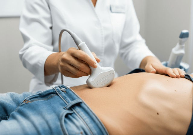
A liver ultrasound is a painless, noninvasive procedure that most people tolerate well. During the exam, you’ll lie on a table while a sonographer applies warm gel to your upper abdomen. The ultrasound probe will press against your skin, creating mild to firm pressure, especially near the ribcage, but it’s rarely uncomfortable. If your liver is tender, let the technician know. You may be asked to hold your breath briefly for clearer images.
Afterward, the gel is wiped off, and there’s no recovery time needed—you can resume activities immediately. Results are analyzed later by a radiologist. Most describe the process as “just pressure” and find it interesting to watch images of their liver in real time.
Can Lifestyle Impact Liver Ultrasound Results?
Your lifestyle can impact liver health and, indirectly, liver ultrasound results. For example:
-
Body weight: Obesity or high BMI can make ultrasounds harder by weakening sound waves. Weight loss may improve image quality.
-
Diet: Large meals or gas-producing foods before a scan can obscure the liver. Following fasting instructions improves results.
-
Alcohol use: Heavy drinking can cause fatty liver or cirrhosis, altering ultrasound visuals. Reducing alcohol and improving diet can normalize imaging.
-
Hydration: While hydration has minimal impact on standard ultrasounds, it can affect stiffness readings in elastography.
-
Activity: Regular exercise and a healthy diet can improve fatty liver, which may show as better imaging over time.
So, while lifestyle might not change the ultrasound physics, it changes your liver’s condition, and a healthier liver makes for a “cleaner” ultrasound result. This is one more motivator to stick to any diet or exercise plans your doctor recommends for liver health. Your efforts can literally be seen on the ultrasound image over time as your liver health improves.
Talking to Your Doctor About Ultrasound Results
After your liver ultrasound, your doctor (primary care physician, gastroenterologist, or hepatologist) will typically share the results with you. It’s important to discuss the findings and next steps to ensure all your questions are answered.
Understand the results: Ask your doctor to explain the report in simple terms. Ultrasounds often use technical phrases, so don’t hesitate to ask, “Does everything look normal?”
If results are normal: Confirm if any follow-up or lifestyle changes are recommended, like diet adjustments for mild fatty liver.
If there’s a finding: Your doctor will outline next steps, such as follow-up imaging or tests, and explain the rationale.
Always ask for clarification, request a copy of the report, and address any concerns or symptoms for a clear plan moving forward.
Monitoring Your Liver Health at Home

While ultrasound is a key part of liver cancer screening, it’s not the only tool that can help you stay proactive about your liver health. Many people are now using at-home liver health tests to get quick insights between doctor visits—especially those managing long-term conditions like fatty liver, hepatitis, or cirrhosis.
Ribbon Checkup is a quick, reliable at-home test that gives you real-time insights into your liver and overall wellness. With just a urine sample and your smartphone, you can track important health markers like bilirubin, urobilinogen, and indicators of liver strain—all in minutes, right from home.
Try Ribbon Checkup today and take the guesswork out of your liver health.
Conclusion
In conclusion, while ultrasound is a valuable first-line tool for detecting liver cancer, especially in high-risk individuals, it’s not infallible. Its noninvasive nature, accessibility, and cost-effectiveness make it ideal for routine screening, but limitations exist, especially for small or hidden tumors. That’s why doctors often use ultrasound alongside other tests like AFP bloodwork, CT scans, or MRIs for a more complete diagnostic picture. Early detection remains the cornerstone of effective treatment, so staying proactive with regular screenings, healthy lifestyle choices, and even at-home liver monitoring can significantly improve outcomes. Always follow up on abnormal results and stay informed—your liver health depends on it.
FAQs: Can Ultrasound Detect Liver Cancer?
1. Can ultrasound detect liver cancer?
Yes, ultrasound is a quick and safe way to detect liver tumors. It's often used first, especially for high-risk patients. However, it may miss very small or hard-to-see tumors.
2. What does liver cancer look like on ultrasound?
It usually appears as a mass that stands out from normal liver tissue—either darker (hypoechoic) or brighter (hyperechoic).
3. Can ultrasound miss liver cancer?
Yes, especially if the tumor is tiny, hidden by bowel gas, or the liver is fatty or scarred.
4. How does ultrasound compare to CT or MRI?
Ultrasound is great for initial screening, but CT and MRI offer more detailed views if something suspicious is found.
5. How often should I get a liver ultrasound?
High-risk individuals should get one every 6 months.
6. What if something shows up on the scan?
Don’t panic—follow-up imaging or blood tests help confirm what it is.
Written by Abel Tamirat, MD
Dr. Abel Tamirat is a licensed General Practitioner and ECFMG-certified international medical graduate with over three years of experience supporting U.S.-based telehealth and primary care practices. As a freelance medical writer and Virtual Clinical Support Specialist, he blends frontline clinical expertise with a passion for health technology and evidence-based content. He is also a contributor to Continuing Medical Education (CME) programs.
Related Resources
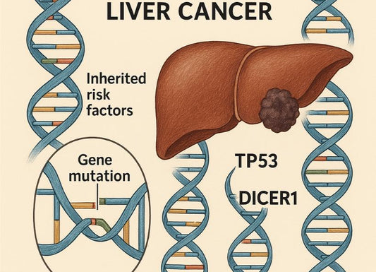
- Is Liver Cancer Hereditary? Genetic Risks and Family History
- Liver Cancer Stages
- What Cancers Cause Elevated Liver Enzymes?
References
Burtka, A. T. (2024, December 31). Liver Ultrasound. Retrieved April 24, 2025, from WebMD website: https://www.webmd.com/fatty-liver-disease/liver-ultrasound
Cafasso, J. (2022, October 20). What Can an Ultrasound Tell You About Liver Cancer? Retrieved April 24, 2025, from Healthline website: https://www.healthline.com/health/cancer/liver-cancer-ultrasound#:~:text=If%20a%20doctor%20or%20healthcare,use%20to%20confirm%20a%20diagnosis
Heald, J., Fetzer, D. T., Rodgers, S., Jain, V., Fung, A., Liu, X., … Marks, R. M. (2024). Patient centered HCC surveillance - complementary roles of ultrasound and CT/MRI. Abdominal Radiology. https://doi.org/10.1007/s00261-024-04678-x
Jacobson, D. R. (2023, April 28). Hepatocellular Carcinoma Imaging: Practice Essentials, Computed Tomography, Magnetic Resonance Imaging. Retrieved April 24, 2025, from Medscape.com website: https://emedicine.medscape.com/article/369226-overview#:~:text=Hepatocellular%20Carcinoma%20Imaging%20,MRI%20can%20detect%20premalignant%20nodules
Liver Cancer Survival Rates | American Cancer Society. (2020). Retrieved April 24, 2025, from Cancer.org website: https://www.cancer.org/cancer/types/liver-cancer/detection-diagnosis-staging/survival-rates.html#:~:text=SEER
RSNA, A. (2024). Abdominal Ultrasound. Retrieved April 24, 2025, from Radiologyinfo.org website: https://www.radiologyinfo.org/en/info/abdominus#:~:text=Ultrasound%20imaging%20is%20a%20noninvasive,blood%20flowing%20through%20blood%20vessels
SINGAL, A., VOLK, M. L., A. WALJEE, R. SALGIA, HIGGINS, P., ROGERS, M., & MARRERO, J. A. (2009). Meta‐analysis: surveillance with ultrasound for early‐stage hepatocellular carcinoma in patients with cirrhosis. Alimentary Pharmacology & Therapeutics, 30(1), 37–47. https://doi.org/10.1111/j.1365-2036.2009.04014.x