What is an Echogenic Liver? Understanding Ultrasound Findings

An echogenic liver appears brighter than usual on ultrasound scans. It is a finding that suggests liver tissue changes often related to fat buildup or scarring. It is not the diagnosis itself. But, it signals your doctor to investigate further to determine the real cause. In most cases, the changes are a result of fatty liver disease (FLD). This condition affects about 32% of the world’s population. The good thing is that many liver issues causing hepatic echogenicity on ultrasound can improve with proper management, especially when caught early.
Key Takeaways
-
An echogenic liver is not a diagnosis or a disease but a sign that there is an issue that is affecting your liver health
-
It looks brighter on ultrasound on ultrasound images, which may be due to increased fat content or tissue scarring
-
Fatty liver disease is the most common culprit, affect about 25% of US adults
-
Many people with echogenic liver do not have symptoms of liver disease, which makes regular checkups important
-
Many cases of fatty liver-related echogenicity can be reversed by a change in lifestyle
-
Early detection increases your chances of completely reversing liver changes
-
Working with your doctor to address the underlying cause is key to improving liver health
What Does An Echogenic Liver Mean?

Your doctor will simply tell you that an echogenic liver is the image of the liver when it is scanned by an ultrasound machine. The finding does not imply disease but merely a sign that there is something that’s affecting your liver tissues.
What is the Hepatic Echogenicity Meaning?

Echogenicity means how much sound waves bounced back during your ultrasound test. Sound waves have different bouncing rates for different organs or tissues in the body. Dense or solid tissues will bounce back more sound waves than those that are fluid-filled or soft tissues.
To put it simply, echogenicity is like the brightness level in a photo. The higher the brightness, increased hepatic echogenicity means having a brighter scan. Lower echogenicity will show up as darker areas. This helps doctors tell different tissues apart. For example, bone will show up brighter than the lungs.
Sound waves travel through your bodily tissues, get bounced off by organs, and return to create images. Your liver does not appear totally dark. It has a certain normal brightness level. But, when your liver shows areas that are brighter than usual, doctors call it “echogenic” or “hyperechoic.”
Why is an Echogenic Liver Significant?
Finding an echogenic liver is significant because it often points to an issue that needs attention. It serves as a warning that something is wrong with your liver health.
Hepatic parenchymal echogenicity in the liver tissue occurs because fat droplets, scarring, and other changes are affecting how sound waves behave as they bounce off your liver. These changes don’t happen in normal and healthy liver.
Since liver problems may get worse in the long run, catching it early in its progression gives you a better chance of completely recovering.
Some liver conditions start without any symptoms (asymptomatic). Your liver may be starting to struggle in its function but you may not know it yet. A simple ultrasound scan can reveal these issues even before you get sick.
But you have to keep in mind that not all echogenic livers mean serious trouble. The finding is a helpful means for the doctor to make further investigation. It helps them find out what really is causing the ultrasound finding. So they can recommend steps to protect or help your liver recover.
Is Fatty Liver Disease the Main Cause?

Yes. Most of the time, fatty liver disease is the common reason for a finding of echogenic liver. About 25% of adults in the United States are affected with FLD. This is equivalent to one in four people having fat accumulation in the liver cells.
Your liver should not store fat in excess. But if 5-10% of fat gets deposited in your liver, you would have a condition called “fatty liver” or in highly medical terms “hepatic steatosis.” The extra fat accumulating in your liver cells will change how the sound waves are read by the machine. And it will look brighter than the normal echogenicity of the liver.
Fatty liver is often linked to lifestyle factors including:
-
Being overweight
-
Not exercising enough
-
Eating too much unhealthy foods with more sugar and fat
It can also happen in people who have type 2 diabetes or those who have long-standing high blood pressure.
But this should not cause any worry because fatty liver from these causes can be reversed with lifestyle changes. Your liver has its inherent capability to regenerate when it gets the right nutrition and support from you.
Can Hepatitis or Cirrhosis Cause an Echogenic Liver?
Yes, both conditions (hepatitis and cirrhosis) can make your liver look brighter than usual on ultrasound. These conditions can affect your liver health in various ways than fatty liver does.
Hepatitis means your liver is inflamed or swollen. It can be a result of viral infections like hepatitis B or C. It can also come from drinking too much alcohol or from autoimmune problems where your body attacks its own liver cells. The inflammation will change the liver’s structure. Hence, it affects how it reflects the sound waves as well.
Cirrhosis, on the other hand, occurs when long-term damage to the liver causes the tissues to scar. The scarring replaces healthy liver cells. The scars don’t function so it does not support the liver’s function at all. Also, the scar tissues are much denser than normal liver tissues so it appears brighter than normal on ultrasound.
Both hepatitis and cirrhosis need medical treatment. The sooner you begin with the treatment, the higher your chances of preventing further damage to the liver.
Are There Other Conditions Linked to Echogenic Liver?
Fatty liver, hepatitis, and cirrhosis are among the most common causes of an echogenic liver. However, other less common causes can also produce the same echogenicity on ultrasound.
-
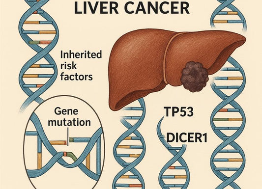
Liver tumors, both benign (non cancerous) and malignant (cancerous), can render some parts of the liver more echogenic. These are masses of growth that have different tissue density than normal liver cells. Which means, they would appear different on ultrasound scans compared to its surrounding tissues.
-
Some metabolic disorders can also affect how your body functions and processes nutrients (metabolism). These are processes that can cause changes in your liver tissues. Examples of these conditions include:
-
Hemochromatosis (too much iron)
-
Wilson’s disease (too much copper)
-
Medications can also cause liver changes, which will also change how it appears on ultrasound scans. This is especially true for long-term use of prescription drugs resulting in increased echogenicity.
-
Rare genetic conditions will also cause abnormal substances to accumulate in the liver, which affects the bounce off rate of sound waves.
However, all of these less common causes will warrant additional testing or investigation, which doctors often recommend. Other testing modalities may be used at this time.

What Are the Symptoms of an Echogenic Liver?
In itself, an echogenic liver doesn’t cause any symptoms at all. The symptoms you may feel are a result of whatever is causing your liver to appear brighter on ultrasound.
Do All Echogenic Livers Cause Symptoms?
Many people with livers that appear bright on ultrasound can have no symptoms at all. This is true when you have fatty liver disease in its early stages. Your liver may store excess fat and show up bright on ultrasound long before it produces symptoms.
The lack of symptoms is why liver problems often go unnoticed for a very long time until they reach a more advanced stage. Your liver works even properly even when it is not completely healthy. It can compensate for any part that’s damaged for a very long time before you feel sick.
There are routine health check-ups that include blood tests or imaging that can help catch these silent liver changes. Finding an echogenic liver by chance during an ultrasound that’s done for another medical reason is actually considered a blessing. It gives you a chance to deal with the underlying issue before it causes significant problems.
There are some people, however, who may have vague symptoms like feeling more tired than usual (easy fatigability) or mild discomfort or dull pain in the upper right side of their abdomen. But these symptoms are non-specific and may be caused by a plethora of conditions. You may even blame it on stress or being overworked.
What Signs Should Prompt a Doctor Visit?
Early liver changes will not cause noticeable symptoms. But while this may be so, certain signs should not be ignored. These signs mean your liver issues are progressing and need medical attention immediately.
So what are these signs?
-
Yellow discoloration of your skin and white portion of your eyes (jaundice) is a serious sign that your liver isn’t working properly anymore. It occurs when your liver can’t process bilirubin, which is a yellow substance naturally found in normal levels in the blood.
-
Persistent pain or a heavy feeling in the upper right part of your abdomen. This sign often signifies that the liver has an ongoing inflammation or enlargement. The discomfort or pain sometimes gets worse after eating fatty foods.
-
Swelling in your legs, ankles, or increasing abdominal girth (circumference). This could mean that your liver is not making enough protein to keep fluids in your blood and usually signify liver failure.
-
Extreme tiredness (fatigability) that does not get better with rest. This could be a sign of liver stress. Since your liver helps in processing nutrients coming from your diet, if it struggles in its function this compromises the energy that you get on a daily basis. If the energy is not enough, you may feel extremely tired and exhausted.
-
Dark urine or pale-colored stools can also signify liver problems. These color changes occur when your liver can’t process waste products effectively.
If you have any of these symptoms, particularly if you already know you have an echogenic liver by ultrasound, seek medical attention immediately.
How Are Symptoms Linked to Underlying Causes
The symptoms you may be experiencing depend on the specific underlying problem that causes your liver to appear brighter than normal on ultrasound. Different liver conditions can cause different symptoms.
Fatty Liver Disease
You may feel tired or have a dull ache on your upper right abdomen, which waxes and wanes. Some people describe a feeling of fullness or discomfort after eating particular foods high in sugar and fat. These symptoms can happen because your liver compresses nearby tissues or organs.
Hepatitis
This condition causes more apparent symptoms such as:
- Fever
- Fatigue
- Stomach pain (sometimes persistent)
There are times that you may lose your appetite or feel nauseated. If it is viral hepatitis, you may feel joint pains or flu-like symptoms, which are basically non-specific.
Cirrhosis
When cirrhosis is at its later stage, it can cause a variety of symptoms occurring along the progression of the disease and the liver function is declining. You may notice the following symptoms:
- Easy bruising or bleeding
- Mental confusion
- Extreme itching (usually because of jaundice)
- Increasing abdominal girth
- Loss of muscle mass even when eating normally
The severity of symptoms usually matches the stage of liver derangement. If the condition is at its early stages, it may cause no symptoms or mild, non-specific ones. However, as the liver problem advances, the symptoms it causes is more concerning.
Remember that symptoms can overlap between liver conditions. This is the reason why proper testing must be done for a more accurate diagnosis.
How is an Echogenic Liver Diagnosed?

Diagnosing echogenic livers involves several steps. It is usually a combination of blood tests, imaging studies, and sometimes taking tissue samples from the liver itself to know what really is causing its appearance on imaging.
Why is Ultrasound the Primary Tool?
Ultrasound is usually the first test when checking for liver health problems. The test is a primary choice because of several reasons, including:
- Safe
- Painless
- Convenient
- Quick
- Does not use radiation
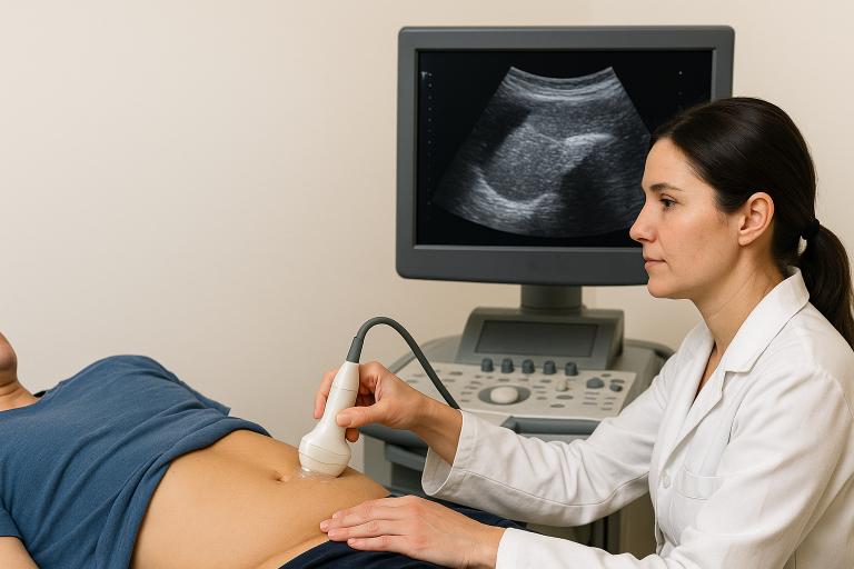
During an ultrasound, the radiologic technician puts a gel on your skin. The technician will then use a device called a transducer and move it over your upper abdomen. The device will send sound waves into your body and capture the waves that bounce back. These bounced off sound waves create images of your liver and other tissues on a screen.
Ultrasound works well for determining the size, shape, and texture of your liver. It will show on the screen if your liver looks brighter than normal, which suggests fatty changes or other issues. It will also detect small lesions or mass or any other abnormal areas that will require further investigation and testing.
The ultrasound will also not require any special preparation from you. There are times when you are required to fast for a few hours before the test, but that’s the most preparation you need to do. The entire test takes only about 15-30 minutes, and you get your results quickly so your doctor can interpret it for you.
Ultrasound tests don’t usually cost as much as MRI (magnetic resonance imaging) or CT (computed tomography) scans do. This means that the test is a practical screening test to check for liver function and health. It is especially true when doctors are screening for common liver issues like fatty liver.
What Other Tests Complement Ultrasound?
While ultrasound can show whether your liver is echogenic or not, it can’t always tell exactly the reason why. That’s where the other tests come into play.
Blood tests can check your liver enzyme levels. The enzyme includes ALT (alanine aminotransferase) or AST (aspartate aminotransferase), which may leak into your bloodstream when a significant amount of liver cells are damaged. Their levels in the blood are usually regulated and will only be within a normal range. So, if there are high levels of these enzymes in the bloodstream, it suggests active liver damage. Other blood tests measure how well the liver makes proteins and clears bilirubin from the blood.
Special blood tests include those that check hepatitis viruses, autoimmune disorders, or genetic liver conditions. These tests can help doctors specify the cause of liver damage.
There are also rare occasions when doctors recommend liver biopsy. This involves taking a tissue sample from your liver with a needle. It is sent for pathology reports, which will show exactly what is happening in your liver cells. It can reveal fat deposits, inflammation, tissue scarring, or other changes.
Newer tests like FibroScan (a specific type of ultrasound that measures liver stiffness and the presence of fat) assess the liver for scarring without a biopsy required. The test is useful for monitoring how liver conditions progress through time.
Sometimes, doctors require further imaging like CT and MRI. These imaging studies give a more detailed picture of your liver. It will demonstrate issues that the ultrasound may have missed.
How Accurate are Ultrasound Findings?
Ultrasound has an accuracy of about 85% when detecting fatty liver when there are 20-30% fat deposits. The accuracy decreases if there are only mild fat deposits. However, it is still the best screening modality for diagnosing the most common cause of echogenic livers—fatty liver.
However, ultrasound has some limitations as a diagnostic tool for fatty liver.
-
It does not accurately detect fat deposits when it is only less than 20%
-
The sensitivity fluctuates from 55-90% when detecting mild fatty liver
-
Cannot detect accurately fatty liver in obese individuals because sound waves have difficulty penetrating thick subcutaneous (under the skin) fat
-
Image quality depends on the skill of the radiologic technician
-
Result depends on the quality of the equipment
-
It is not useful for detecting mild swelling and early-stage scarring. These changes may not affect the echogenicity enough to be noticeable
-
Cannot tell the difference between different types of liver diseases
-
It can only show whether the liver is echogenic but it may not reveal whether the echogenicity is from fat, inflammation, or early scarring.
Because of these limitations, ultrasound is just a starting point. And if it is proven insufficient, doctors request for additional testing and investigation.
How Is Echogenic Liver Treated and Managed?
The treatment and management for echogenic livers depend on the underlying cause. Fortunately, in many cases, it can improve with the right management approach.
Can Lifestyle Changes Reverse Echogenic Livers?
Yes, it can. For many people with FLD, lifestyle changes could be enough in reversing the condition. This is true when the condition is caught early.
Weight loss of about 7% of your body weight to reverse fat buildup and about 10% body weight to reduce scarring.
Dietary changes can also help. You should try to cut back on refined carbs, sugar, and processed foods. These foods increase your risk of developing fatty liver because of the excess fat. You should consider eating more fruits, vegetables, lean proteins, and whole grains.
Regular physical activity is another approach because it helps your body not only in burning fat but also utilize insulin better. Your goal should be about 150 minutes of exercise on a weekly basis, which may include:
-
Brisk walking
-
Swimming
-
Cycling
Even if your goal is not to lose weight, physical activity can still help.
Avoid alcohol and if not at least practice moderation and on occasion. Give your liver a break from processing harmful substances. When you have FLD, avoiding alcohol can help the liver heal faster.
You should also get enough sleep since poor sleeping patterns can worsen insulin resistance, which contributes to FLD. Your goal is to sleep at least 7-8 hours a day.
Stress management techniques like deep breathing exercises and meditation help lower cortisol levels (stress hormone), which can affect your overall health.
These changes, though simple, are steps that could help the liver heal and return to normal within months. With consistent lifestyle changes, the echogenic pattern on ultrasound improves within 6-12 months.
What Medical Treatments are Available?
There are times that lifestyle changes aren’t going to be enough. This is especially true for conditions other than FLD. When this happens, medical treatment is often recommended.
-
Antiviral medications for viral hepatitis
-
Immunosuppressant for autoimmune hepatitis (medications that lower down the activity of your immune system)
-
FLD that are resistant to lifestyle changes may require medications like:
-
Pioglitazone
-
Alcoholic Liver Disease (ALD) requires intervention for substance use
-
Support groups
-
Medications
-
Counselling
-
Management of underlying conditions like diabetes, metabolic syndrome, or high cholesterol
Some people benefit from adding herbal supplements such as milk thistle although scientific evidence is still limited. You should always consult your attending physician prior to starting with any supplement.
It is important to note that these medical treatments only work if combined with lifestyle and dietary changes.
When is Surgery or Advanced Intervention Necessary?
Most cases of echogenic liver, particularly ones caused by FLD, don’t require surgery or any advanced intervention.
Surgery is reserved for complicated cases such as:
-
Severe non-alcoholic steatohepatitis (NASH) especially ones that don’t respond to other treatments like lifestyle changes or weight loss. Bariatric surgery is often recommended.
-
Tumors or growth, particularly hepatocellular carcinoma (HCC), that require procedures for removal
-
Advanced cirrhosis (tissue scarring)
The reason why this is reserved only for complex cases is because FLD rarely progresses once addressed early. This is the reason why early detection is important so that prompt intervention is initiated.
Is an Echogenic Liver Cancerous or Reversible?
You may worry about your long-term health once diagnosed with an echogenic liver. This is the reason why you need to understand every possible outcome.
What is the Prognosis for Echogenic Livers?
The prognosis for echogenic livers depends entirely on what is causing the liver to appear like that on ultrasound. Simple FLD conditions often have good prognosis. For NASH, involving fat deposit with inflammation, the prognosis is still good if caught early in its stages. With proper treatment, the condition could improve or be stabilized.
For cirrhotic livers, the prognosis depends on how much tissue scarring is affecting the liver. Advanced cirrhosis usually doesn't look good but proper treatment can help.
Prognosis will also depend on a number of factors such as:
- Your age
- Treatment response
- Adherence to lifestyle and dietary changes
- How regular your followup is
Conclusion
Liver health is linked to a lot of lifestyle and diet practices. The presence of an echogenic liver—where it appears brighter than usual on ultrasound—is a signal that something is happening to this organ. It is important to determine what causes it so prompt treatment is initiated and potentially reverses the condition.
CTA: For more information and to gain an insight to your liver health from the comfort of your home, visit Ribbon Checkup’s site and get a Ribbon Checkup at-home urine test kit.
Frequently Asked Questions
How to prevent worsening of liver disease?
Active lifestyle, balanced diet, and stress management are enough preventive management strategies that can protect your liver and keep it functioning.
Does Echogenic Liver Lead to Serious Complications?
Yes, if left untreated and ignored, it can lead to serious complications as the disease progresses. However, when properly managed during its early stages, it can repair or prevent the condition from progressing.
Written by Jaclyn P. Leyson-Azuela, RMT, MD, MPH
Jaclyn P. Leyson-Azuela, RMT, MD, MPH, is a licensed General Practitioner and Public Health Expert. She currently serves as a physician in private practice, combining clinical care with her passion for preventive health and community wellness.
References
-
Corey, K. E., & Rinella, M. E. (2016). Medical and surgical treatment options for nonalcoholic steatohepatitis. Digestive Diseases and Sciences, 61(5), 1387–1397. https://doi.org/10.1007/s10620-016-4083-8
-
Fighting Fatty Liver. (2021, September 28). NIH News in Health. https://newsinhealth.nih.gov/2021/10/fighting-fatty-liver
-
Gallacher, J., & McPherson, S. (2018). Practical Diagnosis and Staging of Nonalcoholic Fatty Liver Disease: A Narrative Review. European Medical Journal, 108–118. https://doi.org/10.33590/emj/10314271
-
Gerstenmaier, J. F. (n.d.). Hemochromatosis with hyperdense liver | Radiology Case | Radiopaedia.org. Radiopaedia. https://radiopaedia.org/cases/haemochromatosis-with-hyperdense-liver
-
Gluskin, J. S. (2017). Ultrasound of the liver, biliary tract, and pancreas. Blumgart’s Surgery of the Liver, Biliary Tract and Pancreas, 2-Volume Set, 245-275.e4. https://doi.org/10.1016/b978-0-323-34062-5.00015-7
-
Health Direct. (2019, October 5). Fatty liver. Healthdirect.gov.au; Healthdirect Australia. https://www.healthdirect.gov.au/fatty-liver
-
Hernaez, R., Lazo, M., Bonekamp, S., Kamel, I., Brancati, F. L., Guallar, E., & Clark, J. M. (2011). Diagnostic accuracy and reliability of ultrasonography for the detection of fatty liver: A meta-analysis. Hepatology, 54(3), 1082–1090. https://doi.org/10.1002/hep.24452
-
Hobeika, C., Ronot, M., Beaufrere, A., Paradis, V., Soubrane, O., & Cauchy, F. (2020). Metabolic syndrome and hepatic surgery. Journal of Visceral Surgery, 157(3), 231–238. https://doi.org/10.1016/j.jviscsurg.2019.11.004
-
Johns Hopkins Medicine. (2024). Hepatitis. Www.hopkinsmedicine.org. https://www.hopkinsmedicine.org/health/conditions-and-diseases/hepatitis
-
Lefere, Sander et al.(2022). Intensive Lifestyle Management Improves Steatosis and Fibrosis in Pediatric Nonalcoholic Fatty Liver Disease. Clinical Gastroenterology and Hepatology, Volume 20, Issue 10, 2317 - 2326.e4. https://doi.org/10.1016/j.cgh.2021.11.039
-
Li, Y., Li, B., Jin, F., Ni, J. J., & Wang, J. P. (2025). Assessment of liver involvement in Wilson’s disease with different liver echo patterns based on liver stiffness evaluated on Transient elastography and Sound Touch Viscoelastography. Scientific Reports, 15(1). https://doi.org/10.1038/s41598-025-87859-y
-
NHS. (n.d.). Cirrhosis. Www.nhsinform.scot. https://www.nhsinform.scot/illnesses-and-conditions/stomach-liver-and-gastrointestinal-tract/cirrhosis/
-
Nonalcoholic Fatty Liver Disease (NAFLD). (2025, February 13). American Liver Foundation. https://liverfoundation.org/liver-diseases/fatty-liver-disease/nonalcoholic-fatty-liver-disease-nafld/
-
Practitioners, T. R. A. C. of general. (2013, July). FibroScan and transient elastography. Australian Family Physician. https://www.racgp.org.au/afp/2013/july/fibroscan
-
Teng, M., Cheng Han Ng, Huang, D. Q., En Chan, K., Jun, D., Wen Kwang Lim, Ju Dong Yang, Xiang, E., & Muthiah, M. D. (2022). Global incidence and prevalence of nonalcoholic fatty liver disease. Clinical and Molecular Hepatology, 29(Suppl), S32–S42. https://doi.org/10.3350/cmh.2022.0365
-
Vardan. (2022, December 7). Liver Pain: Major Symptoms Treatment & Causes - PSRI Hospital. PSRI. https://psrihospital.com/liver-pain-major-symptoms-treatment-and-causes/
-
Zhao, Y.-C., Zhao, G.-J., Chen, Z., She, Z.-G., Cai, J., & Li, H. (2020). Nonalcoholic Fatty Liver Disease. Hypertension, 75(2), 275–284. https://doi.org/10.1161/hypertensionaha.119.13419