How Accurate Is Ultrasound for Fatty Liver? Diagnosis, Benefits, and Limitations
 Written By
Blen Shumiye, MD

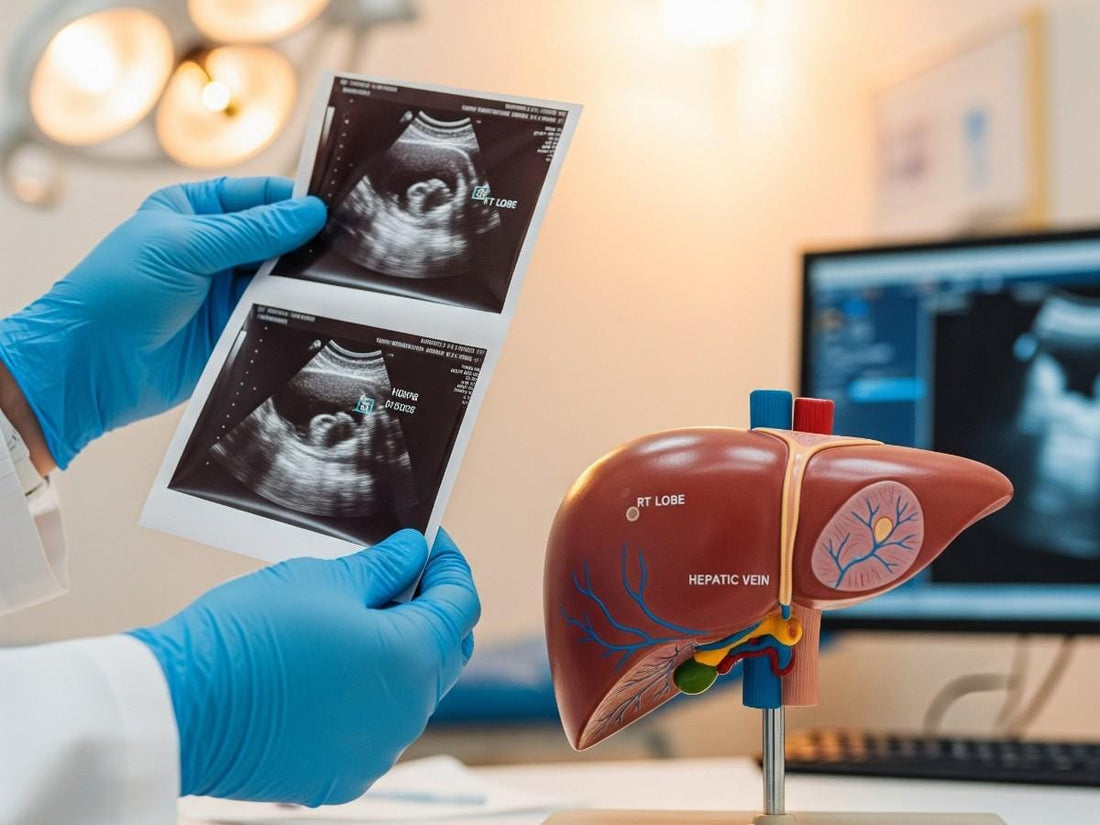
If you’re worried about fatty liver disease, your doctor may recommend an ultrasound as the first step. Ultrasound is quick, painless, and widely available — but how well does it actually detect fat in the liver? The short answer: it’s highly accurate for moderate to severe fatty liver, but it can miss early disease.
This article explores how ultrasound works for fatty liver diagnosis, how accurate it is, what factors affect its reliability, and when additional testing might be needed.
What is fatty liver disease?
Fatty liver disease happens when too much fat builds up in your liver cells, making up more than 5-10% of the liver’s weight. It’s often linked to conditions like obesity or diabetes, but you might not notice symptoms early on. There are two main types:
-
Non-alcoholic fatty liver disease (NAFLD): Now called metabolic dysfunction-associated fatty liver disease (MAFLD), this affects about 25-30% of adults worldwide, with rates as high as 46% in some groups.
-
Alcoholic fatty liver disease (AFLD): Caused by heavy alcohol use, this is less common but still serious.
If untreated, NAFLD can progress to non-alcoholic steatohepatitis (NASH), leading to inflammation, scarring (fibrosis), or even cirrhosis. Early detection is key to preventing these complications, and ultrasound is often the first tool doctors use.
How does ultrasound detect fatty liver?
An ultrasound uses high-frequency sound waves to create images of your liver and nearby organs. The test is done using a handheld device (called a transducer) placed on your abdomen.
When fat builds up in the liver, it changes how sound waves bounce back. On the ultrasound screen, fatty liver appears:
-
Brighter than normal (increased echogenicity)
-
With blurred outlines of blood vessels
-
With poorer visibility of deeper liver tissue
Radiologists use these patterns to decide whether fatty liver is mild, moderate, or severe.
What’s the diagnostic process like?

If your doctor suspects fatty liver—maybe due to high liver enzymes or risk factors like obesity—they’ll likely recommend an ultrasound. Here’s what you can expect:
-
Preparation: You might fast for 4-6 hours to reduce gas in your stomach, which can blur images. Wear loose clothing for comfort.
-
The procedure: A technician applies gel to your abdomen and moves a transducer over it. It’s painless and takes about 15-30 minutes.
-
Results: A radiologist checks the images for signs of fat, like a brighter liver. They’ll also look for other issues, like cysts or tumors.
-
Next steps: If fatty liver is confirmed, you might need blood tests, an MRI, or a biopsy to check for inflammation or scarring.
This quick, non-invasive scan is often your first step toward understanding your liver health.
How accurate is ultrasound for fatty liver detection?

Research shows that ultrasound is highly effective for detecting moderate to severe fat buildup, but not perfect for mild cases.
Key statistics from clinical studies:
-
Sensitivity (correctly detecting disease when it’s present): 80–94% for moderate to severe steatosis.
-
Specificity (correctly ruling out disease when it’s absent): 82–95%.
-
For mild steatosis: Sensitivity drops to about 55–65%.
This means if you have a significant amount of liver fat, ultrasound will likely detect it.
If you have only a small amount, there’s a chance it may be missed.
What are the benefits of using ultrasound for fatty liver?
Ultrasound is often the first test for suspected fatty liver because it’s:
-
Safe: No radiation or injections.
-
Widely available: Offered in most clinics and hospitals.
-
Affordable: Less expensive than MRI or CT scans.
-
Quick: Usually done in under 30 minutes.
-
Repeatable: Can be used to monitor changes over time.
It’s also helpful for spotting other liver or gallbladder issues that may need attention.
For more details checkout ultrasound findings like “echogenic liver”
What affects ultrasound accuracy for fatty liver?
Several factors can influence how well ultrasound detects fat in the liver:
1. Stage of fatty liver
Mild fat accumulation is harder to spot, whereas moderate to severe cases are easier to detect because the brightness change is more obvious.
2. Body weight
Excess abdominal fat can make it harder for sound waves to reach the liver clearly, reducing image quality.
3. Technician skill
Ultrasound is operator-dependent. An experienced sonographer may detect subtle changes that a less experienced one could miss.
4. Machine quality
Modern, high-resolution ultrasound machines provide clearer, more detailed images.
5. Other liver conditions
Scarring (fibrosis), inflammation (hepatitis), or congestion from heart failure can also change how the liver appears on ultrasound.
What can ultrasound tell you and what can’t it tell you?
Ultrasound can:
-
Detect visible fat accumulation in the liver.
-
Estimate severity (mild, moderate, severe).
-
Identify some structural abnormalities like cysts or tumors.
Ultrasound cannot:
-
Identify the exact cause of the fat buildup.
-
Detect inflammation or early scarring without other tests.
-
Measure the exact percentage of fat in the liver.
-
Replace a biopsy for definitive diagnosis.
How does ultrasound compare to other fatty liver tests?
Ultrasound is just one option. Here’s how it stacks up against others:
MRI (Magnetic Resonance Imaging)
-
Most accurate for measuring liver fat, even in mild cases.
-
No radiation, but more expensive and less available.
CT Scan (Computed Tomography)
-
Can detect fat but involves radiation.
-
Less sensitive than MRI for mild fat.
FibroScan (Transient Elastography)
-
Uses ultrasound-based technology to measure both fat and liver stiffness (scarring).
-
Becoming more common in liver clinics.
Blood tests
-
Liver enzymes (ALT, AST) may suggest liver injury but can be normal even with fatty liver.
-
Specialized panels like the NAFLD Fibrosis Score estimate scarring risk, not fat levels.
When should you consider additional testing?
Your doctor might recommend another test if:
-
Your ultrasound is normal but you have strong risk factors (obesity, type 2 diabetes, high cholesterol).
-
Blood tests show abnormal liver function despite a normal ultrasound.
-
You need a precise measurement of liver fat for research or treatment planning.
-
There’s a concern for advanced disease or complications.
In such cases, MRI, FibroScan, or liver biopsy may be used.
Understanding Your Results

After your ultrasound, the report will usually describe your liver appearance as normal, mild fatty infiltration, moderate fatty infiltration, or severe fatty infiltration.
-
Normal: No visible signs of excess fat. If you still have risk factors, your doctor may recommend lifestyle monitoring.
-
Mild: Small amounts of fat are present—may be reversible with early lifestyle changes.
-
Moderate: A significant amount of fat is visible—medical follow-up and more intensive lifestyle changes are often recommended.
-
Severe: High fat content is altering the liver’s appearance—often a sign to investigate for inflammation or fibrosis.
Your doctor may combine these findings with blood tests or additional scans to confirm the diagnosis and assess liver function.
How to improve your liver health after diagnosis?
If your ultrasound shows signs of fatty liver, there’s good news in many cases, especially early on, it’s possible to reverse the damage
Evidence-based ways to help your liver recover:
-
Aim for gradual weight loss – Shedding 7–10% of your body weight can significantly lower liver fat and inflammation.
-
Choose a nourishing, balanced diet – Fill your plate with vegetables, whole grains, lean proteins, and healthy fats.
-
Cut back on added sugars and refined carbs – This helps slow the liver’s fat production.
-
Move more, most days – About 150 minutes of moderate activity a week keeps blood sugar stable and supports fat reduction.
-
Skip the alcohol – Even small amounts can make liver damage worse.
-
Manage other health conditions – Keep diabetes, cholesterol, and blood pressure in check to reduce strain on your liver.
Small, consistent steps can have a big impact. Over time, these habits don’t just improve your liver health; they support your overall energy, metabolism, and long-term well-being.
Consider using our Fatty Liver Diet Plan PDF to naturally support liver recovery.
Final Takeaway:
A liver ultrasound is a safe, simple, and effective way to get a first look at your liver health. Most results either come back normal or show mild changes that can often be improved with straightforward lifestyle adjustments. Even if your scan suggests moderate to severe fatty liver, it’s not the end of the story — many cases can be reversed with the right steps. What matters most is viewing your results alongside your full health picture and working with your doctor on a plan that fits your needs.
In the meantime, you can stay proactive with Ribbon Checkup’s at-home liver health tests and practical guides. These resources make it easier to track your progress, understand your numbers, and take small, steady actions that protect your liver and overall well-being between appointments.
Related resources
● Life Expectancy with Fatty Liver Disease: What You Need to Know
References
Clinic, C. (2023, August 25). A liver ultrasound is a simple and painless way to screen for liver diseases, including cirrhosis, fatty liver, cancer and other lesions. Cleveland Clinic. https://my.clevelandclinic.org/health/diagnostics/liver-ultrasound
Hernaez, R., Lazo, M., Bonekamp, S., Kamel, I., Brancati, F. L., Guallar, E., & Clark, J. M. (2011). Diagnostic accuracy and reliability of ultrasonography for the detection of fatty liver: A meta-analysis. Hepatology, 54(3), 1082–1090. https://doi.org/10.1002/hep.24452
Khov, N. (2014). Bedside ultrasound in the diagnosis of nonalcoholic fatty liver disease. World Journal of Gastroenterology, 20(22), 6821–6821. https://doi.org/10.3748/wjg.v20.i22.6821
Leivas, G., Maraschin, C. K., Blume, C. A., Telo, G. H., Trindade, M. R. M., Trindade, E. N., Diemen, V. V., Cerski, C. T. S., & Schaan, B. D. (2021). Accuracy of ultrasound diagnosis of nonalcoholic fatty liver disease in patients with classes II and III obesity: A pathological image study. Obesity Research & Clinical Practice, 15(5), 461–465. https://doi.org/10.1016/j.orcp.2021.09.002

Dr. Blen is a seasoned medical writer and General Practitioner with over five years of clinical experience. She blends deep medical expertise with a gift for clear, compassionate communication to create evidence-based content that informs and empowers. Her work spans clinical research, patient education, and health journalism, establishing her as a trusted voice in both professional and public health spheres.


