What Is a Liver Hemangioma and Should You Be Worried?


A liver hemangioma (also known as hepatic hemangioma) is a benign cluster of blood vessels in your liver. It rarely causes symptoms. It’s also one of the most common noncancerous liver growths and rarely causes symptoms or needs treatment. For most people, it’s discovered by chance during imaging for another issue. And it almost never leads to cancer or serious complications.
Understanding what it is and how it’s diagnosed can help you stay informed and calm about your liver health. Additionally, knowing when to monitor it and when to seek medical help allows you to stay confident about your health. With smart lifestyle habits, regular checkups, and at-home monitoring tools, you can take a proactive role in keeping your liver strong.
Key Insights
-
A liver hemangioma is a benign cluster of blood vessels, not cancer.
-
Most are discovered incidentally during imaging tests.
-
Hormones and genetics may play small roles in growth.
-
Surgery is only needed in rare, symptomatic cases.
-
Healthy diet and limited alcohol support overall liver health.
-
At-home urine strips can help monitor general liver function.
-
Regular imaging ensures long-term stability and peace of mind.
What Causes a Liver Hemangioma?
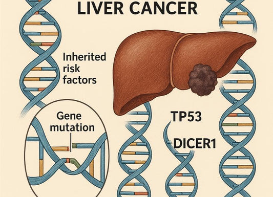
A liver hemangioma forms when small blood vessels in the liver group together in a cluster. There is still no definitive explanation why this happens, which doctors find perplexing. Research suggests that this condition is a mix of genetic, hormonal, and developmental factors, which all play a role.
-
It often appears as a cavernous hemangioma, which means a cluster of larger blood vessels.
-
Most liver hemangiomas are small, less than 4 cms, and cause no harm.
-
Some studies link this growth to estrogen exposure during pregnancy
The condition is usually found in about 60-80% of adults aged 30 to 50. While the exact cause isn’t fully known, it is clear that these growth patterns are not related to cancer or infections of the liver. They’re considered benign vascular tumors, meaning they don’t spread or turn malignant.
Does it run in families?

A liver hemangioma can sometimes appear in multiple family members. However, there is no clear evidence or proof that it is inherited in the traditional genetic way. Some data notes mild familial clustering, which suggests a small hereditary factor is in place although this is not a substantial proof.
However, there is no need to worry if a parent has one, but you can mention it to your doctor during liver screenings.
-
Family history might increase the likelihood even though the risk is low
-
There’s no known gene linked to liver hemangiomas
-
Regular imaging can catch one early if you’re concerned
Can birth control pills or hormones make it grow?
Estrogen may influence liver hemangiomas. That’s why growth can occur during pregnancy or with hormone therapy. Most doctors agree that birth control pills are safe, but large hemangiomas may need monitoring.
-
Estrogen can enlarge vascular tissues
-
Women are affected more often than men
-
If you take oral contraceptives or hormone replacement therapy, tell your healthcare provider
-
Growth doesn’t always require stopping medication but might prompt a follow-up scan
What Are the Signs and Symptoms?
Most people show no symptoms at all. A liver hemangioma is often a silent condition. It may even be discovered accidentally during an ultrasound or CT scan for another reason. However, larger ones (over 5 cm) can cause pressure-related symptoms.
-
You may be feeling pain in the right upper abdomen
-
Some report bloating or fullness even after small meals
-
Nausea and fatigue can also occur when the mass presses nearby organs
Because the liver has no pain nerves inside it, discomfort usually comes from stretching of its outer capsule or compression of surrounding tissue.
When does it become painful?
Pain appears only when the hemangioma grows large or spontaneously bleeds inside itself, which is a rare case. A liver hemangioma larger than 10 cm may cause discomfort or a feeling of fullness.
The pain is characterized as:
-
Pain suddenly feels dull, constant, or located under the ribs on the right
-
Sudden, sharp pain can mean a rupture or internal bleeding, which is an emergency
-
In most cases, mild aching or bloating is harmless but should be checked
Can it cause back or shoulder pain?

Yes, but rarely. If the mass presses on nerves or nearby structures, you may feel pain that radiates to your back or right shoulder. Other liver or gallbladder issues can feel similar, so imaging is the best way to find the cause.
-
Shoulder or upper back pain often mimics gallbladder attacks
-
Pain relief typically comes from managing underlying liver pressure
-
Your doctor may suggest an ultrasound or MRI if pain persists
How Is a Liver Hemangioma Diagnosed?
A liver hemangioma is usually found by accident during an ultrasound, CT scan, or MRI for another issue. Imaging gives doctors a clear picture of blood flow and structure inside your liver.
-
Ultrasound uses sound waves to identify round, hyperechoic (bright) lesions.
-
CT scans can show contrast-filled vascular patterns.
-
MRI is the most accurate and safest for detailed confirmation.
Doctors rarely need to do a biopsy because it can cause bleeding. Instead, radiologists use characteristic imaging signs to confirm the diagnosis confidently.
What does it look like on an ultrasound?
On ultrasound, a hepatic hemangioma looks like a bright, well-defined area in the liver. It reflects sound more strongly than surrounding tissue, which makes it stand out.
-
It’s round or oval and has clear edges.
-
Blood flow appears slow but organized when using Doppler mode.
-
It doesn’t invade nearby structures or spread to other organs.
Ultrasound is often the first test because it’s quick, painless, and uses no radiation.
When is a biopsy needed?
A biopsy is rarely necessary. Doctors might recommend one only if imaging isn’t clear or if another type of liver lesion (like a cyst or adenoma) can’t be ruled out.
-
Biopsies carry a small risk of bleeding because the lesion contains blood vessels.
-
MRI usually provides enough information to confirm a hemangioma safely.
-
If your scan shows an unusual pattern, your hepatologist might suggest additional tests instead of a biopsy.
Imaging Comparison Overview
|
Imaging Method |
Accuracy |
Purpose |
Remarks |
|
Ultrasound |
Moderate |
Initial detection |
Non-invasive, inexpensive |
|
CT-Scan |
High |
Structure and contrast |
Uses radiation |
|
MRI |
Very high |
Blood flow patterns |
Safest for repeated monitoring |
Do Liver Hemangiomas Require Treatment?
Most liver hemangiomas don’t need treatment. If it does, it needs an indication. Doctors usually recommend observation, especially if the lesion is small and symptom-free. You may need periodic imaging every 6 to 12 months.
-
Small hemangiomas (under 4 cm) are left alone
-
Large ones (over 10 cm) or those causing pain might need surgery
-
No medication can shrink or eliminate a hemangioma
Monitoring focuses on stability, ensuring no growth or new symptoms appear.
When is surgery considered?

Surgery is rare. It’s considered only when the hemangioma grows quickly, causes severe pain, or affects nearby organs.
-
Lobectomy removes part of the liver with the lesion.
-
Enucleation removes only the tumor, leaving healthy tissue intact.
-
Surgery carries low risk because the liver regenerates.
Doctors also look at overall liver function before deciding. Most people live full, healthy lives without any operation.
What lifestyle or diet choices help liver health?
A balanced lifestyle supports liver wellness. While diet doesn’t directly shrink a hemangioma, healthy choices keep your liver functioning well.
-
Limit alcohol to protect liver cells.
-
Eat high-fiber foods, lean proteins, and vegetables.
-
Avoid processed sugar and trans fats.
-
Stay hydrated and maintain a healthy weight.
You can also track your liver health at home with urine test strips or wellness kits that measure indicators like bilirubin or protein levels. These tests don’t diagnose hemangiomas but can help monitor overall liver performance and guide when to see a doctor.
When Should You See a Doctor?

You should see a doctor if you develop persistent right upper abdominal pain, fullness after eating, or unexplained fatigue. Most of the time, symptoms are minor, but they deserve evaluation.
-
Schedule an appointment if symptoms last more than a few days.
-
Seek immediate care for sudden severe pain or internal bleeding signs (dizziness, fainting).
-
People with large hemangiomas may need regular follow-up imaging.
Imaging every 6 to 12 months helps ensure the hemangioma stays stable.
Key questions to ask your doctor
-
What size is my hemangioma, and has it changed?
-
Do I need a follow-up scan or additional tests?
-
Are my symptoms related to the hemangioma?
-
Should I avoid hormone medications or birth control?
-
What lifestyle habits best support my liver?
What follow-up imaging might be needed?
Your doctor may recommend:
-
Ultrasound every 6 to 12 months for stability.
-
MRI if the lesion appears larger or irregular.
-
CT scan if you have surgery or new symptoms.
Regular imaging ensures accurate tracking without invasive tests. These follow-ups are preventive, not alarming, and keep you informed about your liver health.
Related Resources
Is F3 Liver Fibrosis Reversible?
Does Exercise Help Liver Cirrhosis?
Quick Summary Box
-
Liver hemangioma is a benign liver tumor made of blood vessels
-
Often, it is less than 4 cm
-
There is usually no symptoms present but may cause pain if large enough
-
The diagnosis is often accidental but may include ultrasound, CT, or MRI
-
Treatment often involves observation, surgery only for severe cases
-
Lifestyle modification includes eating balanced meals, avoid alcohol excess, use at-home wellness tests
-
Call your provider if abdominal pain is persistent or there is rapid growth observed on scans
References
Díez Redondo, P., Velicia Llames, R., & Caro-Patón, A. (2004). Familial hepatic hemangiomas. Gastroenterologia Y Hepatologia, 27(5), 314–316. https://doi.org/10.1016/s0210-5705(03)70467-1
Evans, J., Willyard, C. E., & Sabih, D. E. (2021). Cavernous Hepatic Hemangiomas. PubMed; StatPearls Publishing. https://www.ncbi.nlm.nih.gov/books/NBK470283/
Matos, A. P., Jeon, Y. H., Ramalho, M., AlObaidy, M., & Semelka, R. C. (2016). Lobulated margination of liver hemangiomas: Is this a definitive feature? Clinical Imaging, 40(4), 801–805. https://doi.org/10.1016/j.clinimag.2016.03.005
Miller, V. M., & Duckles, S. P. (2008). Vascular actions of estrogens: functional implications. Pharmacological Reviews, 60(2), 210–241. https://doi.org/10.1124/pr.107.08002
N Bajenaru, Balaban, V., F Săvulescu, I Campeanu, & T Patrascu. (2015). Hepatic hemangioma -review-. Journal of Medicine and Life, 8(Spec Issue), 4. https://pmc.ncbi.nlm.nih.gov/articles/PMC4564031/
Ofer Gemer, Oana Moscovici, Dosoretz, C. L., Linov, L., Peled, R., & Segal, S. (2004). Oral contraceptives and liver hemangioma: a case‐control study. Acta Obstetricia et Gynecologica Scandinavica, 83(12), 1199–1201. https://doi.org/10.1111/j.0001-6349.2004.00551.x
Sandulescu, L., Urhut, C., Sarmis, M., Sandulescu, A.-M., Ciurea, M., Cazacu, S., Iordache, L., Daniela, Cazacu, M., Iordache, S., Sandulescu, & Research, A. (2021). World Journal of Hepatology One stop shop approach for the diagnosis of liver hemangioma Conflict-of-interest statement: Country/Territory of origin: Provenance and peer review: Peer-review model: Single blind Peer-review report’s scientific quality classification Grade A (Excellent): A Grade B (Very good): 0 Grade C (Good): 0 Grade D (Fair): 0 Grade E (Poor): 0. World J Hepatol, 13(12), 1892–1908. https://doi.org/10.4254/wjh.v13.i12.1892
Toro, A., Mahfouz, A.-E., Ardiri, A., Malaguarnera, M., Malaguarnera, G., Loria, F., Bertino, G., & Carlo, I. D. (2014). What is changing in indications and treatment of hepatic hemangiomas. A review. Annals of Hepatology, 13(4), 327–339. https://doi.org/10.1016/s1665-2681(19)30839-7
Wang, A., Chen, H., Huang, Z., Tang, H., Shi, H., Wen, J., Li, Q., Jiang, Y., & Fu, W. (2020). Spontaneous internal hemorrhage of a giant hepatic hemangioma with infection: a case report and literature review. Journal of International Medical Research, 48(12). https://doi.org/10.1177/0300060520976474
Yuranga Weerakkody. (2019). Hepatic hemangioma | Radiology Reference Article | Radiopaedia.org. Radiopaedia.org. https://radiopaedia.org/articles/hepatic-haemangioma-3
Zhang, W., Huang, Z., Ke, C., Wu, C., Zhang, Z., Zhang, B., Chen, Y., Zhang, W., Zhu, P., & Chen, X. (2015). Surgical Treatment of Giant Liver Hemangioma Larger Than 10 cm. Medicine, 94(34), e1420–e1420. https://doi.org/10.1097/md.0000000000001420