What Causes Small Lesions on the Liver? Symptoms, Diagnosis, and Treatment

The liver, a vital organ, performs numerous functions, including detoxifying blood, metabolizing nutrients, and producing essential proteins. Small lesions on the liver are often detected incidentally during imaging tests like ultrasound, CT scans, or MRI. While the discovery of a lesion may raise concerns about cancer or serious liver disease, most small lesions are benign and may not require treatment. However, accurate diagnosis is critical to rule out serious conditions.
This article explores the causes, symptoms, diagnostic methods, and treatment options for small liver lesions, with an emphasis on medical accuracy.
What Are Liver Lesions?
A liver lesion is an abnormal area of tissue in the liver, appearing as a single spot or multiple nodules. Small lesions typically measure less than 2 cm in diameter. Lesions are classified as:
-
Benign (noncancerous): Do not spread and generally pose minimal health risks.
-
Malignant (cancerous): May originate in the liver (primary) or spread from other organs (metastatic).
-
Precancerous or indeterminate: Require monitoring to assess potential progression.
What Causes Small Lesions on the Liver?
Small liver lesions can arise from various causes, ranging from benign to malignant conditions. Below are the primary causes, with medically precise details:
1. Benign Liver Lesions
Hemangiomas
-
The most common benign liver lesion, formed by tangled blood vessels.
-
Typically asymptomatic, small (often <2 cm), and found incidentally during imaging.
-
Rarely cause complications unless large (>5 cm), when they may cause pain or bleeding, necessitating intervention.
Focal Nodular Hyperplasia (FNH)
-
A benign lesion often linked to abnormal blood vessel development in the liver.
-
More common in women aged 20–50, possibly associated with hormonal factors.
-
Typically asymptomatic and stable, rarely requiring treatment unless symptomatic.
Hepatic Adenomas
-
Rare benign tumors linked to prolonged use of estrogen-containing oral contraceptives, anabolic steroids, or metabolic disorders like glycogen storage disease.
-
More common in women of reproductive age.
-
Small adenomas (<5 cm) are often low-risk, but larger ones may bleed or, rarely, transform into hepatocellular carcinoma, requiring surgical evaluation.
2. Cysts
Simple Liver Cysts
-
Fluid-filled sacs, usually benign and asymptomatic.
-
Often solitary and discovered incidentally.
-
Rarely require treatment unless they cause symptoms due to size or compression.
Polycystic Liver Disease
-
A genetic condition causing multiple cysts, often associated with polycystic kidney disease.
-
May remain asymptomatic unless cysts grow large, causing pain or complications.
Hydatid Cysts
-
Caused by parasitic infection (Echinococcus species), typically from contact with contaminated food, water, or animals.
-
Rare in developed countries but serious, requiring medical or surgical intervention.
3. Infectious Causes
Liver Abscesses
-
Caused by bacterial (e.g., pyogenic abscesses), fungal, or parasitic infections (e.g., amoebic abscesses).
-
Small lesions may be early manifestations, often accompanied by systemic symptoms like fever, chills, or right upper quadrant pain.
-
Risk factors include biliary disease, diabetes, or recent abdominal surgery.
Viral Hepatitis
-
Chronic hepatitis B or C can cause inflammation, leading to regenerative nodules or scarring.
-
Over time, these may progress to cirrhosis or increase the risk of hepatocellular carcinoma.
4. Malignant Lesions
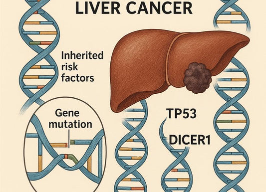
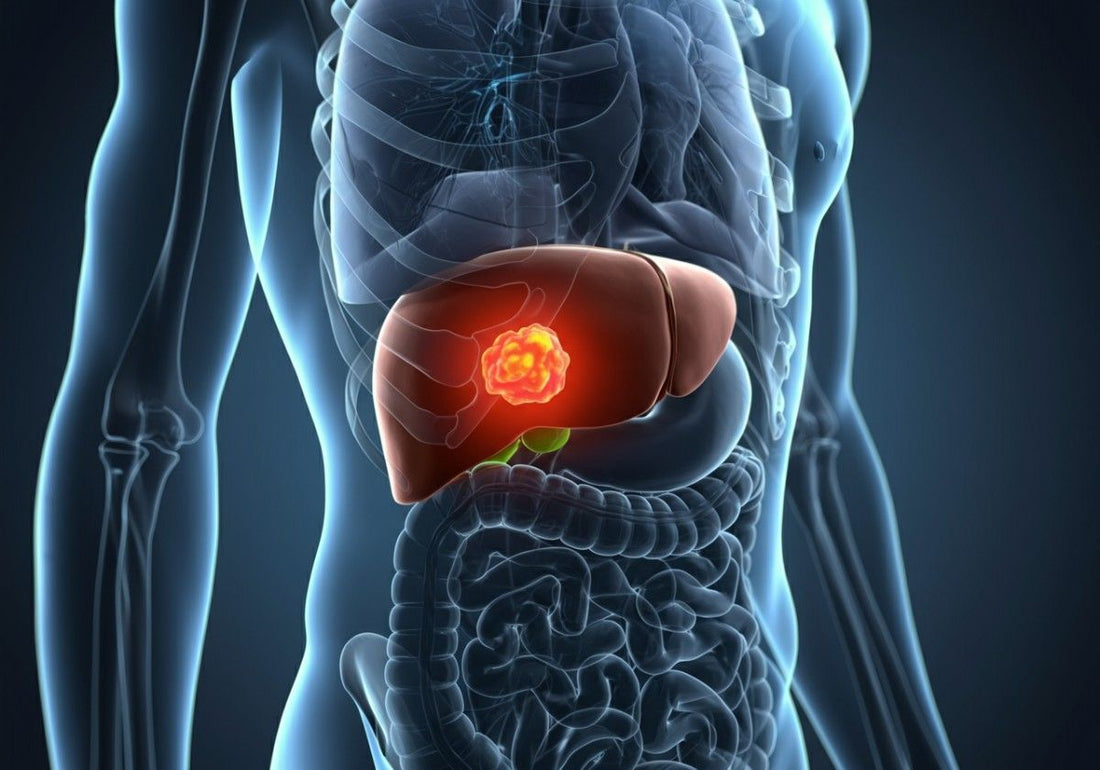
Hepatocellular Carcinoma (HCC)
-
The most common primary liver cancer, often arising in the setting of cirrhosis or chronic hepatitis B/C.
-
Small lesions (<2 cm) may be early HCC, detectable via surveillance in high-risk patients.
-
Risk factors include alcohol-related liver disease, non-alcoholic fatty liver disease (NAFLD), and viral hepatitis.
Cholangiocarcinoma
-
A rare cancer originating in the bile ducts within or outside the liver.
-
Small intrahepatic lesions may be detected early but are challenging to diagnose without advanced imaging or biopsy.
Metastatic Lesions
-
The liver is a common site for metastases from cancers of the colon, breast, lung, pancreas, or stomach.
-
Small lesions may indicate early metastatic spread, often requiring systemic evaluation to identify the primary tumor.
5. Other Causes
Regenerative Nodules
-
Occur in cirrhosis due to repeated liver injury and regeneration.
-
Often benign but may mimic early HCC on imaging, requiring close monitoring.
Vascular Disorders
-
Conditions like Budd-Chiari syndrome or portal vein thrombosis can cause nodular changes mimicking lesions.
Autoimmune or Inflammatory Conditions
-
Autoimmune hepatitis or sarcoidosis may produce granulomas or nodules resembling lesions.
Symptoms of Small Liver Lesions

Most small liver lesions are asymptomatic and discovered incidentally. When symptoms occur, they are often non-specific and include:
-
Mild or dull pain in the right upper quadrant of the abdomen.
-
Fatigue or generalized weakness.
-
Loss of appetite or unintentional weight loss.
-
Fever, particularly with infectious causes like abscesses.
-
Jaundice (yellowing of skin or eyes), typically with malignant or obstructive lesions.
-
Nausea or gastrointestinal discomfort.
These symptoms warrant medical evaluation, as they may indicate underlying liver disease or other conditions.
How Are Small Liver Lesions Diagnosed?
Accurate diagnosis is essential to distinguish benign from malignant lesions. Diagnostic approaches include:
1. Medical History and Physical Exam
-
History: Assesses risk factors such as chronic liver disease, hepatitis, alcohol use, oral contraceptive use, or family history of liver cancer.
-
Physical Exam: Checks for signs of liver disease, such as jaundice, hepatomegaly, or ascites.
2. Blood Tests
-
Liver Function Tests (LFTs): Measure enzymes (ALT, AST, ALP), bilirubin, and albumin to assess liver health. Normal LFTs do not rule out lesions.
-
Viral Hepatitis Serologies: Screen for hepatitis B (HBsAg, anti-HBc) and hepatitis C (anti-HCV, HCV RNA).
-
Tumor Markers: Alpha-fetoprotein (AFP) may be elevated in HCC, though it lacks specificity for small lesions.
3. Imaging Tests
-
Ultrasound: Often the initial test, detecting lesions and guiding further evaluation. Contrast-enhanced ultrasound improves specificity.
-
CT Scan (Multiphasic, Contrast-Enhanced): Provides detailed imaging to characterize lesions based on vascular patterns (e.g., arterial enhancement in HCC).
-
MRI (Liver-Specific Contrast): Highly sensitive for distinguishing benign (e.g., FNH, hemangioma) from malignant lesions.
-
PET Scan: Less commonly used but may help evaluate metastatic disease.
4. Biopsy
-
Reserved for cases where imaging and blood tests are inconclusive.
-
Percutaneous needle biopsy provides a tissue sample for histopathological analysis.
-
Carries a small risk of bleeding or tumor seeding in malignant cases.
Treatment Options for Small Liver Lesions

Treatment depends on the lesion’s type, size, cause, and associated risks:
1. Benign Lesions
-
Hemangiomas, FNH, Simple Cysts: Typically managed with observation via periodic imaging (e.g., every 6–12 months) unless symptomatic or growing.
-
Hepatic Adenomas:
-
Small, stable adenomas may be monitored.
-
Discontinuation of oral contraceptives or steroids is recommended.
-
Large (>5 cm) or symptomatic adenomas may require surgical resection due to risks of rupture or malignant transformation.
2. Infectious Lesions
-
Abscesses: Treated with targeted antibiotics, antifungals, or antiparasitic agents. Percutaneous drainage may be needed for larger abscesses.
-
Hydatid Cysts: Managed with antiparasitic drugs (e.g., albendazole) and sometimes surgical removal to prevent complications.
3. Malignant Lesions
-
Surgical Resection: Preferred for localized HCC or cholangiocarcinoma in patients with adequate liver function.
-
Liver Transplantation: Considered for early-stage HCC in patients with cirrhosis or liver failure, per Milan criteria.
-
Ablation Therapies: Radiofrequency ablation (RFA) or microwave ablation is effective for small HCCs (<3 cm) in non-surgical candidates.
-
Transarterial Chemoembolization (TACE): Delivers chemotherapy directly to the tumor while blocking its blood supply, suitable for intermediate-stage HCC.
-
Systemic Therapies: Targeted therapies (e.g., sorafenib, lenvatinib) or immunotherapy (e.g., nivolumab) are used for advanced HCC or metastatic disease.
4. Monitoring and Supportive Care
-
Regular imaging and blood tests for high-risk patients (e.g., those with cirrhosis or hepatitis) to detect progression early.
-
Management of underlying conditions like viral hepatitis or NAFLD to prevent lesion development.
Prognosis and Outlook
-
Benign Lesions: Excellent prognosis, with minimal impact on life expectancy. Most require only monitoring.
-
Infectious Lesions: Highly treatable with timely intervention, often resolving without long-term complications.
-
Malignant Lesions: Prognosis depends on stage, liver function, and treatment response.
-
Early-stage HCC has a 5-year survival rate of 50–70% with resection or ablation.
-
Advanced disease has poorer outcomes.
When to See a Doctor

Seek medical attention for:
-
Persistent right upper quadrant pain or abdominal swelling.
-
Unexplained weight loss or loss of appetite.
-
Jaundice or dark urine.
-
Fever with abdominal pain, suggesting infection.
-
New liver findings in patients with a history of hepatitis, cirrhosis, or cancer.
Preventing Liver Lesions
While not all lesions are preventable, risk reduction strategies include:
-
Hepatitis B vaccination and safe practices to prevent hepatitis C (e.g., avoiding shared needles).
-
Limiting alcohol intake to prevent cirrhosis.
-
Maintaining a healthy weight to reduce the risk of NAFLD.
-
Using hormonal medications (e.g., oral contraceptives) under medical supervision.
-
Regular screening for high-risk individuals (e.g., those with chronic hepatitis or cirrhosis) via ultrasound and AFP testing.
Key Takeaways
-
Most small liver lesions are benign, often discovered during routine imaging.
-
Causes include hemangiomas, cysts, infections, and cancers, each requiring specific evaluation.
-
Diagnosis relies on imaging, blood tests, and, if needed, biopsy.
-
Treatment ranges from observation for benign lesions to surgery or ablation for malignant ones.
-
Early medical evaluation is critical to determine the lesion’s nature and guide management.
Final Thoughts
Finding a small liver lesion can be unsettling, but most are benign and manageable with proper care. Consult a hepatologist or gastroenterologist for a thorough evaluation, especially if you have risk factors like chronic liver disease or a history of cancer. Early diagnosis and tailored management are key to maintaining liver health.
Take Charge of Your Liver Health
Many liver conditions — including those that cause small liver lesions — may develop silently without obvious symptoms. Early detection is key. The Ribbon Check-Up Urine Test offers a fast, non-invasive way to screen for potential liver and metabolic issues from the comfort of your home.
References
Liver cancer - Symptoms and causes. (2025). Mayo Clinic; https://www.mayoclinic.org/diseases-conditions/liver-cancer/symptoms-causes/syc-20353659
Liver Lesions: What They Are, Types, Symptoms & Causes. (2023, September 7). Cleveland Clinic. https://my.clevelandclinic.org/health/diseases/14628-liver-lesions
Parente, D. B., Araújo, J., Luis, A., Rodrigues, R. S., Perez, R. M., & Marchiori, E. (2018). Fat-containing liver lesions: a pictorial review. Radiologia Brasileira, 51(1), 52–57. https://doi.org/10.1590/0100-3984.2016.0147
Yetman, D. (2022, April 28). What Are Liver Lesions? Healthline; Healthline Media. https://www.healthline.com/health/liver-lesions